Stomach and digestive issues run in my family. While some conditions may be self-inflicted (hello, hot sauce and Thai food), there's also GERD, ulcers and Celiac Disease.
After dealing with an unexplained illness for a few months, I visited my gastroenterologist, who prescribed a barium swallow test with Upper GI Series to see what was going on.1,2
It was my first experience with either of these procedures, so I wasn’t exactly sure what to expect, but the special preparations were a lot easier than a colonoscopy!
Instead of fasting for over a day, the Upper GI Series only required me to avoid eating or drinking anything after midnight on the day of the test. The chalky barium swallow contrast material was a bit hard to get down, but it was still easier than other procedures.
Once the diagnostic radiology procedure highlighting my GI tract was complete, I was ready to go home. No general anesthesia, no hospital gown, chaperone or ride required!
The use of fluoroscopy in radiology and the radiologic technologists who perform it are completely fascinating. Here’s what you need to know.
What is fluoroscopy?
Fluoroscopy is a medical procedure that uses passing X-rays to capture a real-time video of movements inside the body. The images allow healthcare providers to diagnose health problems, see how a patient’s organs, joints and tissue function, and guide through treatments like implants or injections.
To get the best fluoroscopic images, your healthcare provider may inject the dye into your veins, have you swallow it, or apply it to your rectum via a barium enema.
7 Things to know about fluoroscopy in radiology
Fluoroscopy, which uses a fluorescent scope and a continuous beam that pulses through the body, may require a special contrast dye such as barium or an iodine-based contrast material to get the best diagnostic and fluoroscopic views.
While there may be a few small risks, fluoroscopic examinations are typically beneficial for diagnosing issues or guiding certain medical procedures within the body.,
Speak to your healthcare provider for additional details or to answer any questions regarding your fluoroscopy and what the procedure involves. In the meantime, let’s look at some things you should know about this form of diagnostic radiology.
1. Fluoroscopy is a type of X-Ray
Fluoroscopy is an essential diagnostic modality that uses X-rays to provide images of the body in real time.
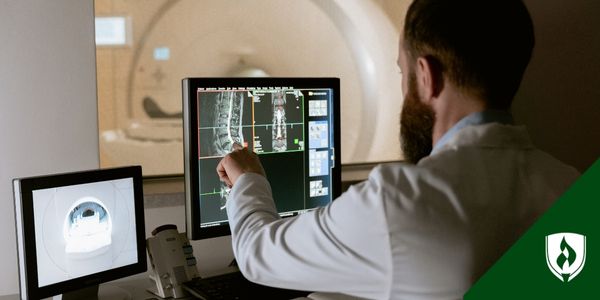
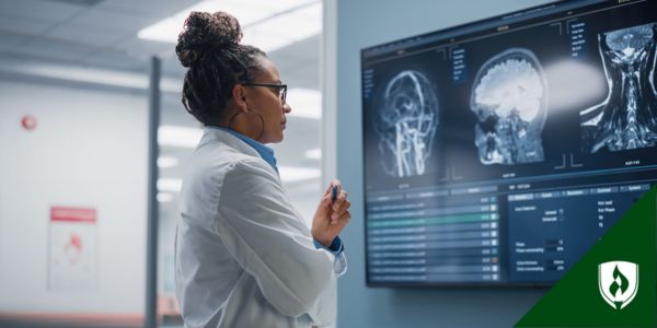
The procedure will typically require imaging guidance and patient positioning from a radiologist or radiologic technologist. These medical professionals use fluoroscopy machines equipped with a fluorescent screen or digital detector to capture images.
2. It's like shooting a video of various body parts
Fluoroscopic procedures can help diagnose problems related to many different parts of the body. Unlike an X-ray that captures a single snapshot, fluoroscopy is like shooting a video where doctors can see how the systems function in real-time.
Some fluoroscopy procedures are outpatient—performed while the patient is awake—while others are same-day, inpatient hospital procedures.
Whether the fluoroscopy procedure involves sedation or anesthesia depends on the body part that needs examination. For example, cardiac catheterization requires sedation since they inspect the coronary arteries and heart and are used alongside catheter placement.
Doctors, radiologists and radiologic technologists may perform fluoroscopy procedures during surgery or stent placement while a patient is under general anesthesia. This is especially true if the type of procedure is complex or painful, if the patient is relatively young or if the patient has disabilities or medical conditions.
Examples of diagnostic fluoroscopy include...
- Angiography: This type of fluoroscopy shows blood flowing in your arteries and blood vessels. It’s used to detect or diagnose narrowing or blockages.
- Arthrography: Used to evaluate joint problems such as tears in cartilage or ligaments.
- Barium swallow: This fluoroscopy procedure helps identify problems in the upper GI tract.
- Barium enema: Examines the colon and rectum to check for abnormalities in the large intestine.
- Small bowel follow-through: A series of fluoroscopy images to evaluate how contrast passes through the small intestine, helping to detect blockages, inflammation or motility issues.
- Cystography: Is a type of fluoroscopy used to detect issues in the bladder.
- Fistulography: A type of fluoroscopy used to evaluate a fistula, or sinus tract, in the skin or soft tissue. A catheter is injected into the sinus or fistula to offer imaging guidance, outlining the path of the tract.
- Hysterosalpingogram: Creates images of the reproductive systems, such as the uterus and fallopian tubes.
- Myelography: Involves injecting contrast around the spinal cord and nerve roots to diagnose herniated discs, spinal stenosis or tumors.
- Orthopedic surgery: A type of fluoroscopy that guides joint replacements and fracture treatments.
- Sniff test: Helps assess how well the diaphragm is functioning, often to diagnose breathing issues.
Providers and radiologic technologists use fluoroscopy for imaging guidance to help with placement of medical devices, injections, or surgical instruments and to see broken bones and fractures. Examples of procedure guidance include...
- Cardiac catheterization: This fluoroscopy procedure uses contrast materials to outline the blood vessels and heart chambers to see how your blood flows through the arteries.
- Catheter insertion or adjustment: Catheters are thin, hollow tubes that deliver or drain fluids into the body. Fluoroscopy helps guide the proper placement of catheters. They can be inserted through the urethra, blood vessels, or bile ducts.
- Orthopedic surgery: A surgeon may use fluoroscopy to help guide certain procedures such as repairing a fractured bone or replacing a joint.
- Stent placement: Stents are small, mesh-like hollow tubes or medical devices that help open narrow or blocked blood vessels. A fluoroscopy treatment can help healthcare providers accurately place stents.
- Biopsies: During a biopsy, fluoroscopy helps guide the biopsy needle to the correct location and ensure an adequate sample of the tissue is removed for accurate testing.
- Vertebroplasty: This procedure stabilizes a broken bone in the spine. Fluoroscopy can help a doctor guide the needle to the right spot, so they can carefully inject cement into the vertebra.
3. Fluoroscopy and radiography serve different medical purposes
While fluoroscopy, radiography and computed tomography (CT) all use X-rays to view parts of the body, they serve different purposes. Radiologic technologists use radiography, or standard X-rays, to capture static, photographic images of internal organs and body systems. It is commonly used to detect a broken bone or fracture, lung ailments or dental issues.
Meanwhile, fluoroscopy provides moving images to allow healthcare providers to observe the motions inside the body. This type of dynamic imaging is even more beneficial when combined with contrast dyes or contrast materials to highlight and provide images of the appropriate body part.
Due to the continuous radiation exposure of fluoroscopy, there is a greater risk of longer-term damage to the body than with traditional X-ray imaging.
4. The procedure may increase radiation exposure risk
While it’s an incredibly useful medical imaging tool, fluoroscopy is not without dangers.
For starters, there are many risks involved with radiation exposure, especially if you’ve recently had certain medical procedures that utilize fluoroscopy, such as barium X-ray treatments or exams. Exposure to radiation can slightly increase the chance of developing cancer later in life.
Plus, exposure to unnecessary radiation can pose a significant threat to pregnant women and cause problems like birth defects and developmental issues.
There is also a small risk of an allergic reaction to contrast dyes, latex and iodine, and taking specific medications could lead to negative interactions. Some patients may experience mild side effects like nausea for a brief period after receiving contrast materials. Liquid containing contrast dye can also impact kidney function, especially risky for those with pre-existing kidney conditions or kidney failure.
Remember that fluoroscopy exams offer critical treatment and diagnostic benefits that typically outweigh the risk of radiation exposure.
When a board-certified radiologist or technologist performs this procedure, a fluoroscopy can be essential and lifesaving too. The FDA regulates all fluoroscopy machines, meaning they meet all federal and state safety standards.
5. Medical imaging preparation varies by exam
Preparation and patient care depend on your procedure, radiation dose, and the contrast agent used during fluoroscopy to better visualize your organs and internal body systems. Your healthcare provider will include instructions for you to follow the days leading up to and on the day of your fluoroscopy.
Special preparations for a fluoroscopy may include...
- Angiography: Since this fluoroscopy test monitors your blood flow and checks for blocked blood vessels, you should check your blood in advance to determine how well it clots. You also shouldn’t take clopidogrel for five days before the procedure or blood thinners within 72 hours before and 24 hours after the test. On the day of your procedure, drink only clear liquids for breakfast and make sure to have someone drive you home.
- Arthrogram: Drink lots of water the day before the fluoroscopy exam. It’s recommended you have a driver take you to and from the fluoroscopy procedure.
- Barium enema: Follow a low fiber diet for a few days before the procedure and then switch to clear liquids like broth, water, or tea the day before your procedure. To ensure your colon is completely clear, you’ll also have to drink a 64-ounce sports drink bottle, Miralax®, and some Dulcolax® tablets. Don’t eat or drink anything on the day of your exam.
- Esophagram: Also known as a barium swallow test, healthcare providers recommend you don’t eat or drink anything for several hours before this fluoroscopy exam and you should not use gum, mints or cigarettes the night before your exam.
- Hysterosalpingogram (HSG): The HSG fluoroscopy exam should be done in the first half of the menstrual cycle (days 1 to 14) to reduce the chance you may be pregnant. Do not have unprotected sex from the start of your cycle until after your exam. Your provider may suggest taking an over-the-counter pain medicine several hours before the test to reduce discomfort.
- Intravenousurogram (IVP): You must fast for four hours and drink lots of water the day before this fluoroscopy exam.
- Myelogram: Do not eat or drink for four hours before your exam. You need to bring someone along to drive you home.
- Upper GI series: The night before your fluoroscopy, do not eat or drink anything after midnight, and do not take your morning medication. Your upper GI tract must be empty for a successful exam.
- Upper GI with small bowel exam: Do not eat or drink anything the night before your exam. Your stomach and intestines must be empty for the best results.
6. The standard process may change based on the procedure
The type of procedure depends on your medical condition, diagnosis, or doctor’s orders. While certain procedures may be performed on an outpatient basis, others may be part of your inpatient hospital stay.
Typically, fluoroscopy follows the same process:
- You might have to remove any clothing or jewelry that could get in the way of the imaging. If you must remove clothing, you'll wear a hospital gown.
- Depending on the type of procedure, you may be given a contrast dye either swallowed, provided via an IV line in your hand or arm, or administered via an enema.
- The patient will assume different positions on an X-ray table. During the procedure, you may have to hold your breath, lie down or move various body parts to improve real-time image quality.
- Certain procedures may require catheter insertion, such as cardiac catheterization, an additional IV line may require proper placement in the elbow, groin, or another other site on the body depending on the procedure.
- Anesthesia or sedation may be required depending on the fluoroscopy examination.
- A special X-ray scanner will capture and produce fluoroscopic images of the body structures and organs.
- Sometimes, a dye contrast may be injected into the IV line to help highlight certain structures or organs.
- For arthrography, fluid in the joint may be withdrawn using a needle before the contrast material is injected. Once the dye is injected, you may have to move the joint to ensure the contrast dye is evenly distributed throughout.
- The length of the procedure depends on the body part being treated or examined, such as the small bowel, colon or spine.
7. These medical procedures use high-tech equipment
A lot has changed with fluoroscopies since the old days. Early fluoroscopies were basic, with an X-ray source and a fluorescent screen, which the patient stood between. Doctors watched as the beams passed through the body.
Today, the fluorescent screen joins forces with an electronic device that amplifies and converts the glowing beam into a video signal that can be presented on a digital monitor or computer screen.
The equipment used in a modern-day fluoroscopy system can provide a continuous series of images produced at a rate from a few images per second to up to 30 images per second. The speed and accuracy of the images produced are a result of the high signal-to-noise ratio (SNR) images, but there are other key components that make these high-tech procedures possible.3
Image receptor
An image receptor—also called an image intensifier or flat panel detector—captures X-rays that pass through your body and turns them into digital images you can see on a computer screen or monitor. Internally, it also brightens and enhances the image so doctors can see more clearly.
Flat-panel detector
An updated version of an image intensifier and image receptor, the flat-panel detector offers increased sensitivity while reducing a patient’s radiation dose. The FPD consists of a matrix of photodiodes that detect and convert X-ray photons into an electronic signal. Contrast ratio and temporal resolution have also improved, reducing the blurring motion and covering a wider latitude.
Image intensifier
Similar to the image receptor and flat-panel detector, the image intensifier is an electronic device that transforms the X-ray beam intensity pattern into an image that can be picked up by a camera shown on a screen.
Patient table and pad
A patient table is an adjustable X-ray table where the patient lies down during the exam. It’s equipped with gears and motors to move in different positions to capture the best view during X-ray and fluoroscopy procedures. Foam pads are sometimes placed between the patient and the table to add a layer of comfort while adjusting the patient’s position.
X-ray tube
An X-ray tube, located in the head of the fluoroscopy machine, generates X-rays when a high-voltage current passes through. It’s essentially a vacuum tube in which electrons speed up and then collide with a target, generating X-ray photons.
X-ray generator
A generator is like an X-ray tube in the sense that it’s the source of the high tube potential that powers the X-ray tube.
Contrast agents
A contrast agent or contrast material refers to a substance used during a fluoroscopy, magnetic resonance imaging (MRI), or computed tomography (CT) scan to highlight specific parts of the body. They change the way ultrasound waves, X-rays, and other types of radiation pass through the body. Depending on the type of test, the contrast agent may be given to the patient rectally, orally or intravenously. There is a low risk of an allergic reaction.
Beam filtration
Many fluoroscopy systems use special aluminum or copper beam-shaping filters to reduce radiation exposure, while producing high-contrast images of the body part. Beam filtration may be manual or automatic, switching between low or higher dose modes during the procedure. Additionally, many fluoroscopy systems use wedge filters to reduce brightness and radiation dose.
Collimator
This device shapes the X-ray beam and controls the shape and size of the area being captured. It’s made of adjustable metal blades or shutters that open or close to control where the beams go to maintain field of vision and minimize exposure during a fluoroscopy.
Anti-scatter grid
When a patient has a fluoroscopy, some of the rays may scatter as they pass through the body, blurring the image and reducing quality. An anti-scatter grid is a device used in fluoroscopy and X-rays to reduce scatter radiation and improve image quality.
Ionizing radiation
Fluoroscopy is ionizing radiation used by healthcare professionals and radiologic technologists in medical imaging procedures which removes electrons from atoms and molecules in the body, allowing different tissues to absorb them at various rates.
A common form is X-rays, according to the CDC, which can penetrate our body and reveal pictures of our bones.4 Examples of this type of radiation at work include computed tomography (CT) scans and magnetic resonance imaging (MRI).
Interested in exploring a career as a radiologic technologist?
If you’d like to learn more about fluoroscopy and radiology, then you should consider a career as a radiologic technologist! It’s a fascinating and rewarding career, offering the unique opportunity to help people and make a difference in their lives.
To learn more about the field, check out our article What Does a Radiologic Technologist Do? An Inside Look at the Job.
Miralax® is a registered trademark of Bayer HealthCare LLC
Dulcolax® is a registered trademark of A. Natterman & Cie. GmbH
1The material contained in this blog is for informational purposes only and is not to be interpreted as advice or guidance
2Barium Swallow Test, Cedars-Sinai Healthcare system, https://www.cedars-sinai.org/programs/imaging-center/exams/gastrointestinal-radiology/barium-swallow.html
3Signal-to-noise ratio (radiography), Joachim Feger on 2 Apr 2020. https://radiopaedia.org/articles/signal-to-noise-ratio-radiography?lang=us
4About Ionizing Radiation, February 19, 2024, CDC https://www.cdc.gov/radiation-health/about/ionizing-radiation.html